

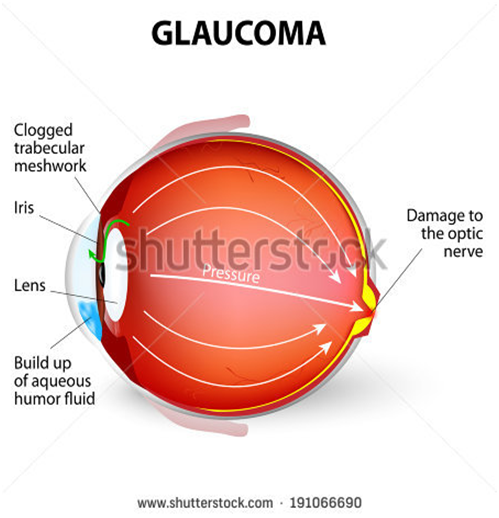
Glaucoma refers to a group of disorders that lead to damage to the optic nerve, due to the increased fluid pressure in the eye. The optic nerve carries visual information from the eye to the brain .Damage to the optic nerve causes vision loss.
TYPES: there are 4 types of glaucoma-
1- Open angle or chronic glaucoma
2- Close angle or acute glaucoma
3- Congenital glaucoma
4- Secondary glaucoma
ETIOLOGY AND RISK FACTOR – Family history of glaucoma ,older age, diabetes, cardiovascular disease, migraine, myopia, eye trauma, prolong use of corticosteroids
PATHOPHYSIOLOGY- Due to the etiology and risk factors (emotional stress , illness, long term corticosteroids)
Turns structural alterations in the aqueous outflow system
It leads to functional alteration condition such as increased intra ocular pressure
Than due to the alteration of the function ,the optic nerve damage
When the arophy of an optic nerve
Visual loss.
CLINICAL MANIFESTATIONS-
1 chronic or open angle glaucoma
Visual impairement
Blurring visin
Mildly aching eyes
2 close or acute angle glaucoma
Severe eye pain
Red eye deceased or cloudy visin
Swelling of the eye
Pupil doesn’t react to light
Headache
Nausea and vomiting
3 congenital glaucoma
Tearing
Sensitivity to light
Red eye
Enlarge ment of one eye or baoth eye
Cloudyness of the front of the eye
DIAGNOSTIC TEST –
1 Tonometry- this test is used to measures the intra ocular pressure.
Normal intra ocular pressure ranges from 10 to 21 mmhg.
2 slit lamp examination –this test is used for see the effect of glaucoma on the anterior eye structure including the cornea , iris, and lens.
3 opthalamoscopy – this test is for facilitates visualization of fundus.
MEDICAL MANAGEMENT-
Epinephrine , mannitol(osmotic diuretic) , isosorbide (to reduce the effect of blood glucose level)
SURGICAL MANAGE MENT-
Trabeculectomy
Iridectomy
Filtering procedure
NURSING DIAGNOSIS-
RISK FOR BLINDNESS FROM EXTREMELY HIGH INTRA OCCULAR PRESSURE CAN BE ADDERESSED AS A COLLABORATIVE PROBLEMS.
RISK FOR INEFFECTIVE THERAPEUTIC REGIMEN MANAGEMENT RELATED TO COMPLEX MEDICATION SHEDULE.
ANXIETY RELATED TO FEAR OF BLIND NESS NAD HOSPITALIZATION.
PAIN REALATED TO SURGERY.
NUTRITION DEFICIENCY RELATED TO SURGERY.
Keywords:
Increased intra occular pressure,
Optic nerve damage,
Open angle glaucoma(chronic),
Close angle glaucoma(acute),
Congenital,
Secondary,
Management (medical and nursing),
DESCRIPTION-
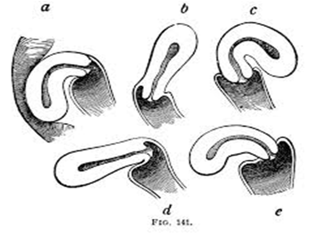
The uterus is not a fixed organ. Minor variation in position in any direction occurs constantly with changes in posture, with straining, with full bladder or loaded rectum. Only when the uterus rests habitually in a position beyond the limit of normal variation, should it be called displacement.
RETROVERSION-
Definition-retroversion is the term used when the long axes of the corpus and cervix are in line and the whole organ turns backwards in relation to the long axis of the birth canal.
RETROFLEXION-
Retroflexion signifies a bending backwards of the corpus on the cervix at the level of internal os. The two conditions are usually present together and are loosely called retroversion or retrodisplacement.
DEGREES-
Conventionally, three degrees are-
FIRST DEGREE- The fundus is vertical and pointing towards the sacral promontory.
SECOND DEGREE- The fundus lies in the sacral hollow but not below the internal os.
THIRD DEGREE- The fundus lies below the level of the internal os.
CAUSES-
1]DEVELOPMENTAL
2]ACQUIRED
DEVELOPMENTAL
Retrodisplacement is quite common in fetuses and young children. Due to developmental defect, there is lack of tone of the uterine muscles. The infantile position is retained. This is often associated with short vagina with shallow anterior vaginal fornix.
ACQUIRED
1] PUERPERAL-
The stretched ligaments caused by childbirth fail to keep the uterus in its normal position. A subinvoluted bulky uterus aggravates the condition.
2] PROLAPSE
Retroversion is usually implicated in the pathophysiology of prolapsed which is mechanically caused by traction following cystocele .
3] TUMOR
Fibroid, either in the anterior or posterior wall produces heaviness of the uterus and hence it falls behind.
4] PELVIC ADHESION
Adhesions either inflammatory, operative or due to pelvic endometriosis pull the uterus posteriorly.
INCIDENCE
Retroversion is present in about 15-20% of normal women.
CLINICAL PRESENTION
1] Chronic premenstrual pelvic pain
2] Backache
3] Dyspareunia
4] Infertility
DIAGNOSIS
1] Bimanual examination
2] Speculum examination
3] Rectal Examination
The retrodisplacement may be confused with hard faecal mass in the rectum, small fibroid on the posterior wall of the uterus and small ovarian cyst in the pouch of Douglas.
PREVENTION
1] To empty the bladder regularly
2] To increase the tone of the pelvic muscles by regular exercise.
3] To encourage lying in prone position for half to one hour once or twice daily between 2 to 4 weeks postpartum.
COORECTIVE TREATMENT
1] PESSARY
2] SURGICAL
PESSARY-Use of pessary is almost obsolete in present day obstetric/gynecological practice.
Usually HODGE- SMITH Pessary is used. The pessary acts by stretching the uterosacral ligaments so as pull the cervix backwords.
SURGICAL TREATMENT
The principle of surgical correction is ventro suspension of the uterus by plicating the round ligaments of both the sides extra peritoneally to the under surface of the anterior rectus sheath .
This will pull the uterus forwards and maintains it permanently.
Haemoglobin Formation.
Description: – It is the red, oxygen carrying pigment in the RBCs. It consist of the protein globin united with the pigment haem.
The Iron in the haem the ferrous form. Each ferrous combines loosely and reversibly with one molecules of oxygen.
There are 4 haem to the one molecules of haemoglobin, contains 4 iron atoms and can carry 4 molecules of oxygen.
Molecular weight of haemoglobin is 68,000.
SOME IMPORTANT DEFINITION:-
1. Oxyhaemoglobin:- Haemoglobin react with oxygen to form oxyhaemoglobin and is represented as HbO2.
2. Carbamino –Haemoglobin:- Carbon dioxide reacts with haemoglobin to form carbamino-haemoglobin.
3. Reduced Haemoglobin:- Haemoglobin from which oxygen has removed.
4. Carboxy Heamoglobin:- Carbon Monoxide reacts with Haemoglobin to form carboxy haemoglobin.
5. Methaemoglobin:- When either reduced or oxygenated haemoglobin is exposed to various drugs to various drugs or oxidizing agents ,the ferrous is oxidized to ferric form and the compound is called methaemoglobin.
NORMAL VALUES:
1. At birth 23gm/dl, because RBC count s more.
2. At the end of 3 month 10.5gm/dl
3. Adults:
Males :- 14-18gm/dl
FemaleS:- 12-15.5gm/dl
FUNCTIONS:-
1. Transport of oxygen from lungs to tissues.
2. Transport of CO2 from the tissues to the lungs.
3. It acts as an excellent acid base buffer,being a protein.
SYNTHESIS:-
Synthesis of haemoglobin requires the provision of nutrients e.g. proteins ,vitamins.
It only takes place in the developing RBCs.
FACTORS CONTROLLING HAEMOGLOBIN FORMATION:-
1. Role of proteins:- It helps in globin formation.
2. Role of minerals:- IRON – It helps in formation of haem. COPPER, COBALT AND CALCIUM – helps in the absorption ,mobilization and utilization of iron.
VARIETIES OF HAEMOGLOBIN:-
Adult haemoglobin
Foetal heamoglobin:- Its structure is same as of HBA, except that the beta chains are replaced by gamma
JISHU BAIJU
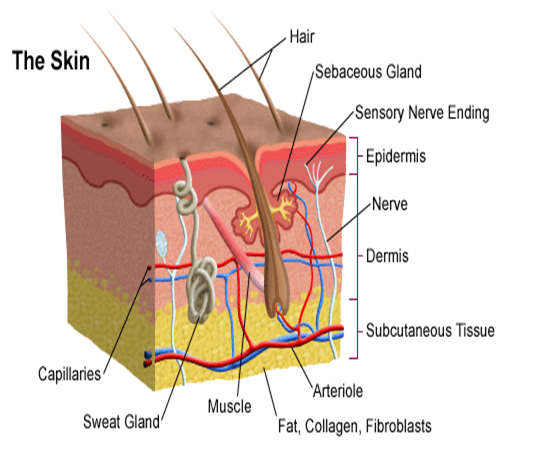
Description: SKIN: – The skin is the continuation of the membranous lining the body openings. It contains glands, hair, nails and consists of two layers.
1. Epidermis
2. Dermis
EPIDERMIS:- The outer skin:-
It is the most superficial layer of the skin and made up of stratified epithelium. It is thickest on the palms of the hands and soles of feet. It contains no blood vessels and nerve cells and its nutrition is derived from the capillaries of the dermis. The Epidermis consists of five layers:
1. Stratum corneum:- It is the most superficial layer and it is made up of dead and highly Keratinised cells.
2. Stratum lucidum:- It is made up of clear non granulated cells.
3. Stratum granulosum:- It consist of granular cells and is about three cells layer thick.
4. Stratum spinosum:- It is 3-5 cells thick layers. The individual cells have a prickly appearance due to the fact that cytoplasmic processes come out from each cell to meet their fellow cytoplasmic strands.
5. Stratum germinativum :- It is the deepest layer of epidermis, made up of columnar cells which give rise to the new cells. It also contains melanocytes which synthesize a pigment called melanin.
DERMIS:- It is a thick and tough elastic layer of connective tissues which supports the hair.
1. Hair :- The hair develops from the hair follicles located in the epidermis.The part of the hair above is the shaft and the remainder ,the root. The colour of the hair depends on the amount of melanin pigment.
The Arrector pilli muscle is attached to the hair follicle.
2. Sebacious glands :- They develops from the hair follicles and usually open into it but in the face they may open into the exterior directly. They are most numerous in the skin of the scalp.
The secretion of the sebaceous glands is rich in oily substances that keep the hair soft.
3. Sweat glands: – They are basically a highly coiled tubular structure in the deeper part of the dermis. Sweat is synthesized by the coiled portion of the sweat glands from water, salt urea and other waste products. Two types of sweat glands are recognized: – Apocrine sweat glands and Ecrine sweat glands.
FUNCTIONS:-
1. PROTECTIVE FUNCTION:- Ultra violet rays of sun i.e. light rays less than 360 um in wavelength can cause damage to the skin producing sun burn and cancer of skin , called ultra violet radiation injury.
2. SENSE PERCEPTION:- Skin provides a sensory covering over the entire body.It contain nerve ending as well as various receptors for general sensation such as touch, pain, pressure and temperature.
3. TEMPERATURE REGULATION:- The different organs of the body by way of metabolism generate large quantities of heat which reaches the skin via blood .
4. WATER BALANCE:- The first layer of epidermis is such a membrane that it does not allow water to pass in and out.
5. Skin helps to eliminate waste products such as urea in the sweat.
JISHU BAIJU
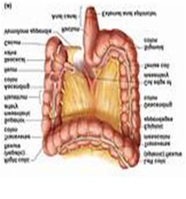
Description:
– Mucosal surface of colon is smooth i.e. there are no villi on the Mucosa.
– Simple tubular glands are abundant which secrete Mucus.
– Wall of the colon is usually folded into sacs by contraction.
– The longitudinal smooth muscle layer is collected into the three distinct bands called taenia coli.
Movement :
Two main types of waves of contraction are seen in the large intestine
A. Segmental Contraction : –
They are of two types.
Type I contraction :
These are small amplitude waves which aid mixing of the contents of the colon.
Type II Contraction :
These are larger pressure waves which aid mixing of the contents of the colon and facilitate absorption by exposing more of the contents of the colon.
B. Peristaltic contractions :
They are also of two types :
1. Type III contraction : These are very small pressure wave of prolonged duration. They propel the contents towards the rectum.
2. Type IV : contraction : This is also called Mass Action contraction. These are simultaneous contraction over a large portion of the colon.
Function :
These contractions propel contents from caecal region towards the rectum. They are the predominant contraction force during defecation.
Both segmental & peristaltic contraction occur infrequently but are particularly evident following a meal. Thus, distension of the stomach by the food initiates contraction of the rectum and frequently a desire to defecate callled Gastrocolic Reflex.
Absorption & Secretion :
– The main function of the colon is absorption of water.
Main sites of water absorption are caecum and ascending colon.
– No digestive enzymes are secreted in the colon.
– At birth, the colon is sterile but the colonic bacterial flora becomes established early in life.
Some of these are beneficial and others harmful.
The Faecus :
1. The faecus are derived :-
(i) Mainly from the intestinal secretions
(ii) Partly from the ingested food.
2. If vegetables and ground cereals are excluded from the diet, the faces have fairly constant composition.
3. The brown colour of stools is due to pigment formed from the bile pigments by the intestinal bacteria.
4. On an average fat intake of 100 gm/day, 5-6 gm/day is normally post in faecus where as on an average protein intake of 100 gm/day 1.5 gm/day nitrogen is normally lost in faecus.
5. Dietary Fibres :
In humans there is no appreciable digestion of dietary fibres e.g. cellulose, hemcellulose and ligin etc. due to absence of certain micro-organism in GIT which break down these substances. Therefore, ingested cellulose passes out unchanged and substances which are enclosed in cellulose wall escape digestion and absorption.
Jishu Baiju